Detecting breast cancer in mammogram images using a Convolutional Neural Network (CNN) based Deep Learning model.
-
Conventional detection of breast cancer tumours in mammograms is a complex task that involves clinical examination, radiological imaging, and pathology testing and can only be accomplished by domain experts.
-
These methods can be automated using machine learning which relies on numerical attributes. However, feature detection continues to be to be a manual task and can be further overcome using deep learning approaches.
-
Hence, in my study I have focused on breast cancer detection using deep learning.
-
Breast cancer is one of the most common cancer diseases among women. According to the International Agency for Research on Cancer (IARC), it accounts for 22.9% of invasive cancers and 13.7% of cancer-related deaths in women across the globe.
-
Therefore, early and accurate diagnosis is critical as it can minimise the death rate and improve the prognosis of breast cancer.
-
Since manual diagnosis is a tedious task, there exists an imperative need to automate breast cancer detection using deep learning.
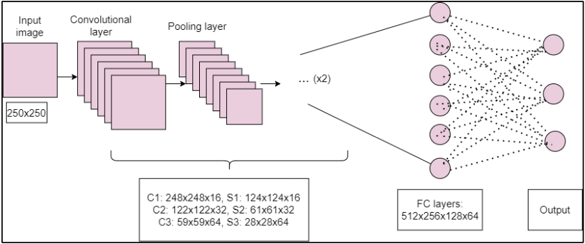
The Convolutional Neural Network (CNN) used comprises of the following architecture:
- Three convolutional layers with 16, 32 and 64 filters respectively, using the Rectified Linear Units (ReLU) activation function.
- A max pooling layer after every convolutional layer with a pool size of 2.
- After the series of convolutional and pooling layers, there exists four fully connected layers with 512, 256, 128 and 64 neurons respectively.
- Finally, in the CNN there exists an output layer with three neurons corresponding to three classes- Normal, Benign and Cancer.
The aforementioned CNN architecture can be graphically seen below.
-
At the onset, it was observed that most of the images in the image datasets were not in a standardised format. The images contained noise in terms of annotations and artefacts which were introduced due to malfunctioning of the device and/or poor illumination.
-
Proceeding further, the annotated images were filtered out and the artefacts were eliminated by creating a uniform black background in a subset of the images.
-
These processes were manually performed (leading to fewer samples) and in the future I would like to automate them.
-
Initially built a Logistic Regression based Machine Learning model using the numerical dataset with ~96% accuracy (More details about the datasets used can be found in the end).
-
To further automate feature detection and extraction process in mammogram images, built a CNN based Deep Learning model and increased its accuracy from 66.6% to 88.8%
-
My current work focused on automating the detection and classification of breast cancer in mammogram images using deep learning approaches.
-
Due to the unavailablity of large and noise-free datasets, the experimentation that was based on a limited number of samples, manual noise-removal methods and different combinations of optimisers achieved a maximum accuracy of 88.8%
-
In the future, I would like to automate the noise-removal process using sophisticated image processing techniques based on Median filters and Gaussian filters to name a few.
-
The automation would lead to the generation of a large number of samples which would better train the CNN and ultimately lead to a better accuracy.
- Breast Cancer Wisconsin (Diagnostic): 357 Benign, 212 Malignant samples.
- The Mini–MIAS Database of Mammograms: 56 Benign, 52 Malignant samples.
- Mini-DDSM: 2728 Normal, 3360 Benign, 3596 Cancer samples.
Hi, I am Arjun Kohli. I'm currently pursuing a Computer Science major at Krea University.
This project was built under the guidance of professor Pavan Kumar Perepu at Krea University. I also had constant help from my peer Ashutosh Verma (Computer Science major, Krea University) throughout the course of the project.